Keratoconus is a progressive condition that can compromise your sight. Left untreated, it may cause significant visual impairment and affect the quality of your life.
However, prompt diagnosis and effective treatments like corneal cross-linking can help delay or halt keratoconus. Keep reading to learn more about when you should consider corneal cross-linking for keratoconus.
What is Keratoconus?
Keratoconus is a condition in which your cornea, the transparent dome-shaped tissue at the front of your eye, gradually thins and weakens, bulging forward like a cone. The cone shape distorts your vision and makes it difficult to see clearly.
Keratoconus typically affects both eyes, but one eye can be more severe than the other.
What are the Symptoms of Keratoconus?

In the early stages of keratoconus, you may experience symptoms such as:
- Eye redness
- Slightly blurred vision
- Halos around bright lights
- Double vision in one eye
- Straight lines appearing wavy or bent
- Increased sensitivity to glare and light
In the later stages of keratoconus, symptoms can include:
- Increased astigmatism or nearsightedness
- More distorted or blurry vision
- Being unable to wear contact lenses
What is Corneal Cross-Linking?
Corneal cross-linking is a procedure used to treat keratoconus. It involves using ultraviolet (UV) light and vitamin B2 eye drops to reinforce bonds between collagen fibers in your cornea and keep it from bulging further.
For this procedure, your ophthalmologist at Herschel LASIK and Cataract Institute will numb your eye with anesthetic eye drops. You may also be given medication to help you relax.
They’ll then gently remove the epithelium or outer layer of the cornea and apply riboflavin or vitamin B2 eye drops. It will take approximately 30 minutes for the eye drops to penetrate deeper into your cornea.
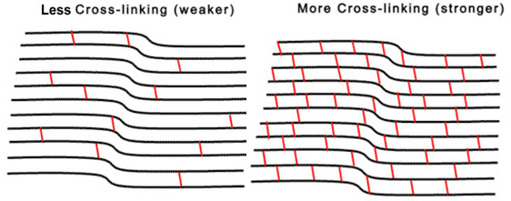
After that, they’ll apply focused UV light to your cornea for about 30 minutes. The UV light activates the riboflavin, setting off a chemical reaction that helps form new bonds between collagen fibers in your cornea to make it more rigid.
Collagen strengthens and maintains the shape of your cornea.
The bonds formed shorten and thicken collagen fibers, resulting in a stronger cornea.
Your ophthalmologist will then apply steroid and antibiotic eye drops to prevent infection and reduce inflammation. A contact lens will be placed over your eye as a bandage as your cornea heals.
You can go home the same day as your procedure, but you’ll need a friend or loved one to take you until you’re cleared to resume driving. You’ll continue to use the steroid and antibiotic drops to ensure smooth healing for 2 to 3 weeks.
When Should You Consider Corneal Cross-Linking?
You can often correct your vision with glasses or specialized contact lenses in the early stages of keratoconus. However, if your vision continues to worsen, you may want to consider corneal cross-linking.
Corneal cross-linking can slow or stop keratoconus from progressing, preserve your vision, and prevent the need for a corneal transplant, which is a more invasive surgery. Generally, you should meet the following criteria to be eligible for corneal cross-linking:
- You have mild to moderate keratoconus
- You do not have corneal scarring
- You do not have certain autoimmune conditions
- You are not pregnant or nursing
- You do not have dry eye syndrome
- You do not have a history of ocular herpes
The only way to know if you’re a suitable candidate for corneal cross-linking is to visit your ophthalmologist at Herschel LASIK and Cataract Institute for a complete assessment. During your appointment, Dr. Herschel will evaluate various factors to determine if corneal cross-linking could be a promising treatment option for you.
How Can You Improve Your Corneal Cross-Linking Outcome?

Combining corneal cross-linking with other treatments can help further improve vision and slow keratoconus progression. For certain patients, corneal cross-linking can be combined with treatments such as:
Topography-Guided PRK
Over time, the irregular shape of a cornea with keratoconus can make it challenging to see clearly with glasses or contact lenses. Topography-guided PRK (TG-PK) is an advanced form of PRK eye surgery that offers more precise vision correction.
It leverages corneal topography technology to create three-dimensional maps of the surface of your cornea. The topography data obtained is then combined with your refractive error and used to guide an excimer laser, precisely reshaping your cone-shaped cornea to a more normal curvature.
This improves the overall quality of your vision and can help correct refractive errors like nearsightedness, farsightedness, and astigmatism.
Intacs® Corneal Implants
Intacs® are semi-circular plastic rings that are surgically implanted into your cornea to flatten its cone shape. The new shape reduces the effects of keratoconus and improves your vision.
Conductive Keratoplasty
Conductive keratoplasty is a procedure in which high-frequency radio waves are used to shrink targeted areas on your cornea, improving the cornea’s curvature.
Save Your Sight with the Right Keratoconus Treatment
At Herschel LASIK and Cataract Institute, we have extensive experience treating patients with keratoconus and offer a wide range of effective treatments, including corneal cross-linking. After a comprehensive eye exam and a thorough assessment, Dr. Herschel can create the best treatment plan to help preserve your vision.
Do you want to slow or stop the progression of keratoconus and protect your vision? Schedule your appointment at Herschel LASIK and Cataract Institute in Orlando, FL, today to determine if corneal cross-linking is right for you.